EN
EN
 TH
TH CN
CN
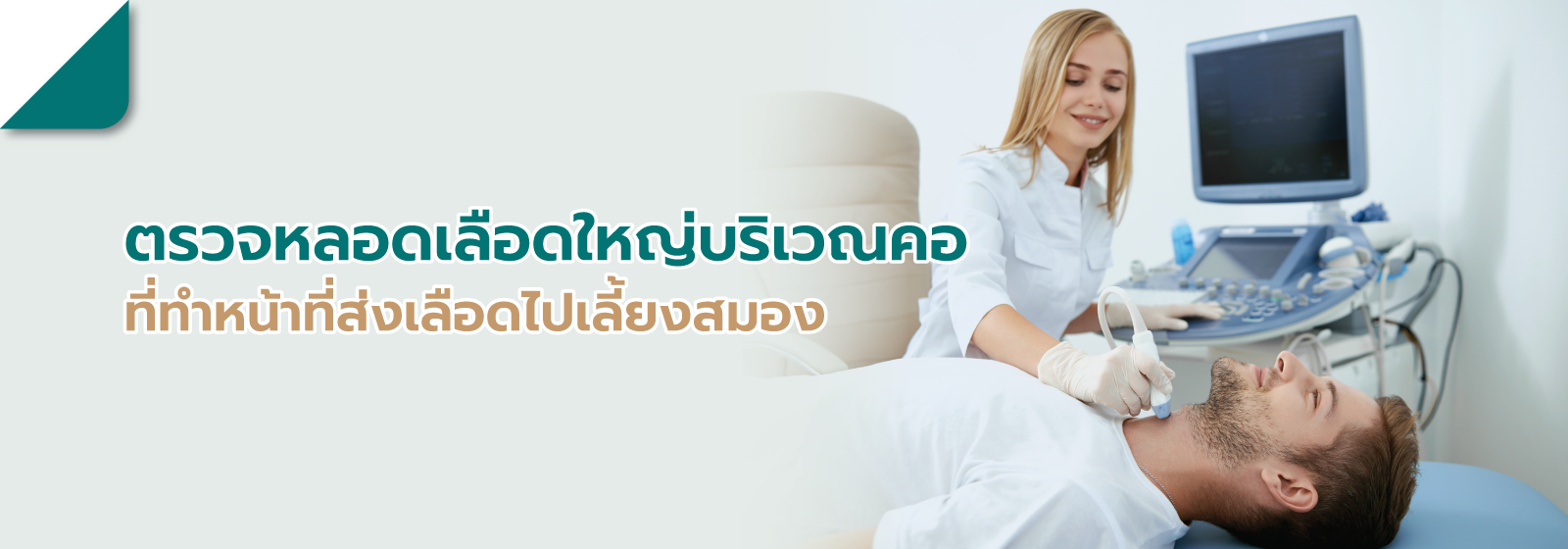
Carotid Duplex Ultrasound


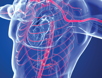
Carotid Duplex Ultrasound is a diagnostic procedure used to evaluate the large blood vessels in the neck that supply blood to the brain. It examines both the front (Carotid Artery) and back (Vertebral Artery & Basilar Artery) systems of the blood vessels using high-frequency sound waves. This ultrasound technique allows for the assessment of both the structure and the characteristics of blood flow within the blood vessels, helping to assess the risk of future narrowing of the arteries, which could lead to conditions like stroke.
This machine uses high-frequency sound waves to examine the neck area while lying down or sitting. The person being examined doesn't need to prepare anything special. After the examination, they can continue with their daily activities as usual.
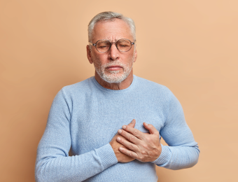
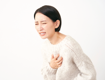
Symptoms that may indicate blood vessel narrowing:
- Carotid Bruit sound detected in the neck during a physical examination by a doctor
- Detection of a moving lump in the neck
- Temporary loss of vision
- Ischemic stroke due to carotid artery narrowing
- Transient ischemic attack
- Monitoring of blood vessel condition supplying the brain after neck artery surgery
- Experiencing dizziness and unsteady gait
Who should be screened:
- Patients with any type of cerebrovascular disease
- Patients in whom Carotid Bruit sound or a moving lump in the neck is detected during examination
- Patients with carotid artery narrowing supplying the brain, both those who have undergone surgery and those who have not
- Individuals with risk factors for cerebrovascular disease:
- Hypertension
- High cholesterol
- Diabetes
- Smoking
- Family history of cerebrovascular disease
- Patients with coronary artery disease
- Patients with peripheral vascular disease
- Patients with aortic aneurysm
This machine uses high-frequency sound waves to examine the neck area while lying down or sitting. The person being examined doesn't need to prepare anything special. After the examination, they can continue with their daily activities as usual.
01 Mar, 2020